Role of contrast enhanced MRI in differentiating tumefactive demyelinating lesions and gliomas
DESCRIPTION
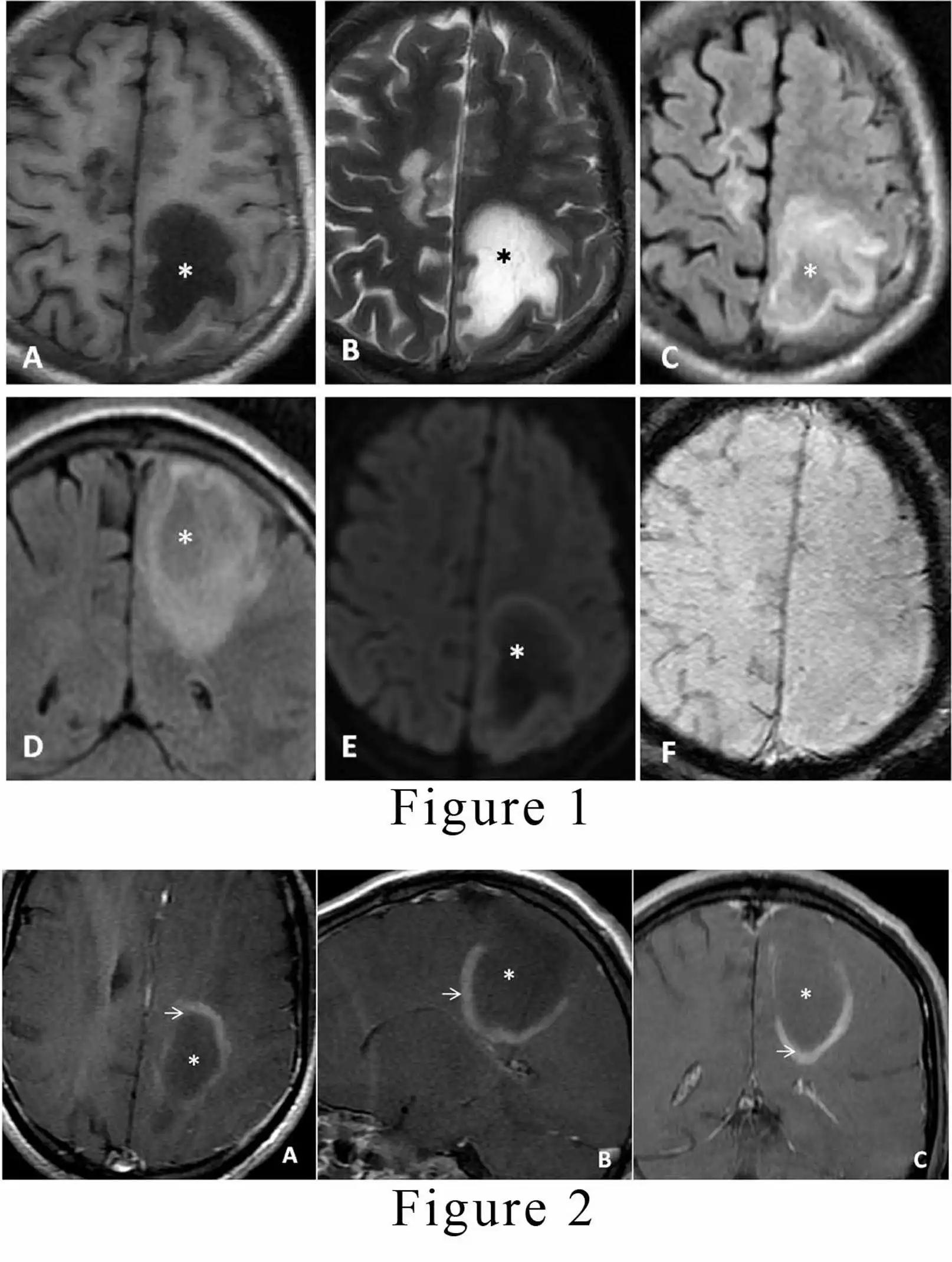
Tumefactive demyelinating lesions (TDLs) are large lesions, usually greater than 2 cm. They typically occur as a solitary lesion or as few separate lesions. [1,2] There is a slight predominance in women with a mean age of 37 years. These lesions usually do not originate as post-infectious or post-vaccination status and do not progress to multiple sclerosis.[1] The patient usually presents with focal neurological deficit, aphasia or seizures. There is typical involvement of supratentorial white matter sometimes extending to involve cortical gray matter. On contrast-enhanced magnetic resonance imaging (MRI), there is an open ring type of enhancement.[2,3] The incomplete portion of the ring is on the gray matter side of the lesion. The enhancing part of the ring represents the acute demyelinating process, whereas the non-enhancing part represents the chronic phase of the inflammatory phase. A 42-year male presented to our institute with a history of blurred vision, paresthesia and motor weakness on right side since one month. There was no history of any similar episode, vaccination or respiratory tract infection. He had a 2/6 vision in the right eye with a normal B-scan and fundal examination on ophthalmic evaluation. The routine blood and serological investigations were unremarkable. For further evaluation, he was advised an MRI scan of the brain, which revealed two abnormal intensity lesions in bilateral frontoparietal lobes; the right-sided lesion measured 27 x 9 mm, and the left lesion measured 52 x 25 mm. The lesions were hypointense on the T1 weighted image, and predominantly hyperintense on both T2 and FLAIR weighted images.[Figure 1A, 1B, 1C, and 1D] There was only minimal surrounding oedema present without any significant mass effect over the adjacent brain. No diffusion restriction or blooming was observed in the lesions on diffusion-weighted images and susceptibility-weighted images, respectively.[Figure 1E, and 1F] The suspicion of glioma and demyelinating disease was raised on the basis of a non- enhanced MRI. For further analysis, contrast-enhanced MRI was advised. On post-contrast sequence, the lesions demonstrated peripheral arc-like enhancement giving a characteristic open/incomplete ring appearance.[Figure 2A, 2B, and 2C] The diagnosis of tumefactive demyelination was made based on clinical history and MRI features. The patient was started on standard steroid treatment, and the patient recovered completely from his visual symptoms and paresis. Presently he is on follow up with our hospital and is doing fine.
CONCLUSION
-
Tumefactive demyelinating lesions are greater than two cm which are often confused with gliomas.These lesions usually involve white matter portion of the brain and sometimes extend to the gray matter portion of the brain.
-
Contrast enhanced MRI helps in differentiating these lesions from gliomas by showing an open/ incomplete type of ring enhancement.
-
Accurate diagnosis of these lesions can prevent the patient from undergoing invasive interventions .
LEGENDS:
-
Figure 1. Unenhanced magnetic resonance imaging (MRI) brain (A, B, C, and D) axial T1 weighted imaging (T1WI), T2WI, Fluid attenuation inversion recovery sequences (FLAIR), and coronal plane FLAIR respectively; demonstrate an irregular abnormal signal intensity lesion with perilesional edema (asterisk) following fluid signals involving left high parietal region predominantly involving white matter. Figure 1 (A, B, and C) axial images shows similar lesionin right fronto-parietal region. (E and F, axial planes) The lesion in left parietal region does not demonstrate diffusion restriction or blooming on diffusion-weighted imaging (DWI) and susceptibility-weighted imaging (SWI) respectively.
-
Figure 2. T1WI, contrast-enhanced MRI (A, B, and C) axial, sagittal, and coronal planes respectively; demonstrate peripheral incomplete ring enhancement (white arrow) with the non-enhancing central area (asterisk) of the lesion. Open portion of the ring enhancemnet is on gray matter side.
REFERENCES
-
Dagher AP, Smirniotopoulos J. Tumefactive demyelinating lesions. Neuroradiology 1996; 38: 560–565.
-
2.Kepes JJ. Large focal tumor-like demyelinating lesions of the brain: intermediate entity between multiple sclerosis and acute disseminated encephalomyelitis? a study of 31 patients. Ann Neurol 1993; 33: 18–27. Google Scholartumefactive demyelination.
-
Masdeu JC, Moreira J, Trasi S, Visintainer P, Cavaliere R, Grundman M. The open ring: a new imaging sign in demyelinating disease. J Neuroimaging 1996; 6:104 –107.