UCOR WEB SERIES
Forthcoming event | Please don't miss out
Past Events
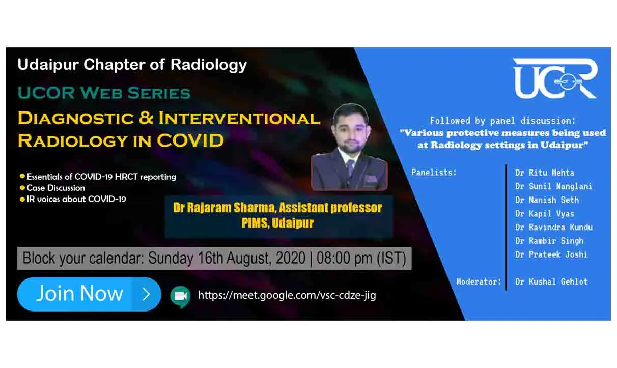
First Trimester ultrasound Scan(11 to 14 weeks)
The first trimester ultrasound scan, which typically occurs between 11 to 14 weeks of pregnancy, is a crucial diagnostic tool used to assess the health of the fetus. In this interactive case-based discussion, we will go beyond examining the nuchal translucency (NT) or nasal bone (NB) and explore other important factors that can be evaluated during this scan.
DR. DEEPAK YADAV
M.D. Radio-diagnosis
Consultant Radiologist & Fetal Medicine Consultant at Dr. Deepak’s Imaging Centre, Rewari (Haryana).
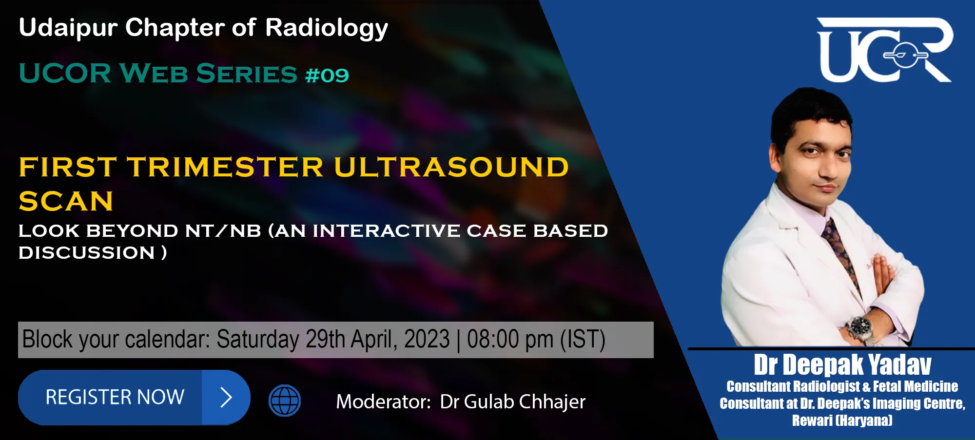
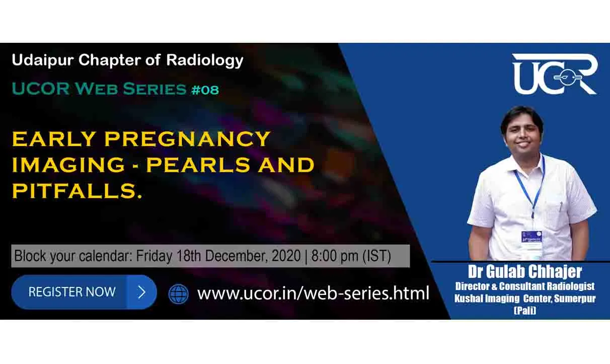
Early Pregnancy Imaging - Pearls and Pitfalls
This talk focuses on 4-10 week period of the fetal development and highlights the awareness of normal appearance and assessment of gestational sac, yolk sac & embryo from 4 weeks gestational age onwards, criteria and terminology of viable and nonviable pregnancy, when to diagnose early pregnancy failure, how to determine chorionicity and amnionicity in early twin gestations and finally decision making in difficult situations of early pregnancy.
Dr Gulab Chhajer
Director & Consultant Radiologist, Kushal Imaging center, Sumerpur (Pali)
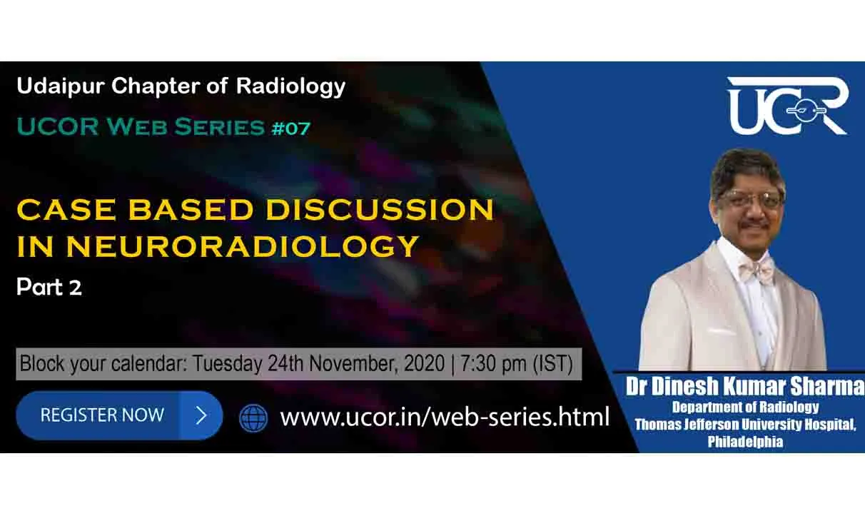
Case based discussion in Neuroradiology
Part 2
"Eyes don't see what your mind doesn't know" To enhance your knowledge and approach towards cases UCOR web series is back with Case Based Discussion in Neuroradiology-Part by Dr Dinesh Kumar Sharma on10th of November' 2020
Dr Dinesh Kumar Sharma
Department of Radiology Thomas Jefferson University Hospital, Philadelphia
Case based discussion in Neuroradiology
Part 1
"Eyes don't see what your mind doesn't know" To enhance your knowledge and approach towards cases UCOR web series is back with Case Based Discussion in Neuroradiology-Part by Dr Dinesh Kumar Sharma on10th of November' 2020
Dr Dinesh Kumar Sharma
Department of Radiology Thomas Jefferson University Hospital, Philadelphia
MRI of the Shoulder
Part 2: How to report common conditions in shoulder?
MRI of the shoulder is one of a frequent examinations faced in daily radiological practice. The bony structures of the shoulder are proximal humerus, the glenoid, coracoid and acromion process and the distal clavicle. The glenohumeral and acromioclavicular joint are two important joints in the shoulder. The Supraspinatus, Infraspinatus, Teres minor and Subscapularis tendons forms the rotator cuff. Common pathologies in the shoulder joint includes: Rotator cuff tear, arthropathies, shoulder impingement, calcific tendinitis, adhesive capsulitis, shoulder instability, labral tears etc
Dr Sanjay Desai
Consultant Radiologist Deenanath Mangeshkar Hospital, Pune
MRI of the Shoulder
Part 1: Anatomy and Basics
MRI of the shoulder is one of a frequent examinations faced in daily radiological practice. The bony structures of the shoulder are proximal humerus, the glenoid, coracoid and acromion process and the distal clavicle. The glenohumeral and acromioclavicular joint are two important joints in the shoulder. The Supraspinatus, Infraspinatus, Teres minor and Subscapularis tendons forms the rotator cuff. Common pathologies in the shoulder joint includes: Rotator cuff tear, arthropathies, shoulder impingement, calcific tendinitis, adhesive capsulitis, shoulder instability, labral tears etc
Dr Sanjay Desai
Consultant Radiologist Deenanath Mangeshkar Hospital, Pune
Ultrasonography in cystic lesion of scrotum.
Scrotal cystic lesions are cause of scrotal mass & painful scrotum. Most of the cystic lesions are benign & can arise from any scrotal content. Ultrasound imaging is a modality of choice for characterization and diagnosis of the same , & hence, unnecessary surgery can be avoided . At the same time Doppler application is helpful to ascertain vascular lesions with confidence. The presentation will be very useful for residents as it will include normal sonoanatomy & variety of case series, congenital to acquired, presenting as cystic mass or scrotal fluid collection, in our day to day clinical ultrasound practice.
Dr Subhash Tailor
Director & Senior consultant Radiologist, Subhash Ultrasound Imaging Center, Bhilwara
Imaging of fetal Urinary Tract
Imaging of Fetal Urinary Tract evaluation is one of the essential part of routine as well as anomaly scan.
Among all Urinary tract (UT) dilation is most common abnormality & sonographically identified in 1–2% of fetuses. The lack of correlation between prenatal and postnatal US findings and final urologic diagnosis has been problematic, in large measure because of a lack of consensus and uniformity in defining and classifying UT dilation. Consequently, there is a need for a unified classification system with an accepted standard terminology for the diagnosis and management of prenatal and postnatal urinary tract dilatation.
Dr Gulab Chhajer
Director & Consultant Radiologist, Kushal Imaging center, Sumerpur (Pali)
Diagnostic & Interventional Radiology in COVID
Discussed the salient features of Covid-19 pneumonia on HRCT and highlighted the importance of CORADS and CT severity score. According to him physicians are more relying on the HRCT findings to treat the patient, rather than viral load or clinical findings.
Dr Rajaram Sharma
Assistant professor PIMS, Udaipur
